I originally wrote this as a printed handout in January 2020. The text is unchanged apart from small typo fixes; the original PDF is embedded at the bottom.
How does the brain keep track of time?
If you are interested in learning some neurobiology, this section is the place to start. Otherwise, feel free to skip ahead to the more interesting stuff that you can actually apply to your day to day life. Thank you for your interest!
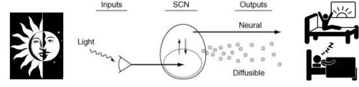
Shorthand summary: there are specialized cells in your eye that respond to light and connect directly to an area of your brain that is called the Suprachiasmatic Nucleus (the body’s ‘master clock’).
The main way in which our body tells time is through light. This is done through light input directly into cells in the retina called rods and cones, which then sends a signal to intermediary cells we call interneurons. Just like a road connecting two destinations, or a charging cable connecting your phone to your computer, interneurons act as a pathway for the input from the rods and cones to connect directly to another type of neuron referred to as ganglion cells. The axons (long nerve fibers, also cable-like in their own way) of these ganglion cells then exit the eye as the optic nerve, which then becomes the optic tract. This tract sends the input we receive from our surroundings to the visual cortex, which is the area of our brain that creates the pictures that we see.
Not all of these ganglion cells’ axons go straight to the visual area though, since about 5% of them, connected to cells called ipRGCs, have only one job: to detect the presence of light in a broad area. ipRGCs are especially light-sensitive compared to the other cells found in the eye, which is why they are called intrinsically photosensitive retinal ganglion cells. The reason why they are so sensitive is because they contain a special photopigment (light-sensitive molecules) called melanopsin in conjunction with receiving photonic input from the interneurons. This means that if for some reason the melanopsin doesn’t work, then there is a backup means for receiving light input.
These cells are very sensitive to light at the ~480nm wavelength, which we perceive as blue light, and artificial light sources, such as white LEDs, have a lot of blue light.
Instead of connecting to the visual cortex however, ipRGCs plug into a very small brain region that neuroscientists refer to as the SCN (suprachiasmatic nucleus), which sits on top of the optic tract closer to the base of your brain. Using blind people as an example, who can either be circadian blind or non-circadian blind, we can see this separation very clearly. Non-circadian blind people have no conscious vision, but are still able to distinguish between light and dark and sleep on time, while circadian-blind people have impaired ipRGCs and have a very difficult time maintaining a 24hr schedule on their own.
Shorthand summary: blue light suppresses our brain’s ability to produce melatonin, which is required to signal the onset of sleep.
Within the SCN, there are about 20,000 neurons (referred to as ‘clock cells’), each of which cycles within a 24hr timeframe. How this cycle works is through what we call a feedback loop: very similar to how thermometers work in how they only actively circulate warm or cold air based on temperature readings and then stop once the room reaches the desired setting, clock genes are self-regulating in the same way.
Now, remember the axons that we were just talking about that go straight to the SCN? At the end of these axons are what are called axon terminals, and if we extend our charging cable metaphor further, we can think of the axon as the cable itself, and the terminals as the actual plug. Similar to how plugs are a specific shape, axon terminals, depending on what the function of the neuron they’re attached to serve, will release a specific chemical that has a certain type of effect on whatever they are ‘plugging into’. In this example, the axon terminals that are connected to the ipRGCs will release two molecules: glutamate and PACAP. Without going into too much detail, these two molecules (mainly glutamate) trigger the 24hr clock gene cycle within each clock cell, and the average of all of these cells expressing this cycle at the same time is what determines our expressed circadian rhythm.
Glutamate, depending on the time of release has the ability to cause what is called either a phase advance, or phase delay in your sleep patterns (please see phase response curve to light). Fibers from the hypothalamus (where the SCN is) then go down to the spinal cord, and makes its way through several brain structures before ultimately reaching the pineal gland. The pineal gland is responsible in this circuit for the secretion and synthesis of melatonin, which is formed from serotonin. Therefore the pineal gland receives its information about the time of day from the SCN, whose genetic regulatory cycle responds to the presence of light.
Therefore, depending on when bright light reaches our eyes we can manipulate our circadian rhythm.
Why are there early birds and night owls?
Chronotype, a term created by circadian researchers, refers to the behavioural expression of a person’s circadian rhythm as a result of their biological clocks. In normal terms, it’s basically the typical time a given individual would naturally fall asleep and wake up. At the more extreme ends of this spectrum, there are people who have a delayed sleep onset relative to the general population (what we call ‘night owls’), as well as an advanced sleep onset (‘early birds’). In chronobiology they are referred to as Delayed Sleep Phase Disorder (DSPD) and Advanced Sleep Phase Disorder (ASPD) respectively.
Chronotype is the result of underlying neural mechanisms, particularly regarded as a genetic predisposition, however artificial light exposure can interact with your biological rhythm in such a way that it can exacerbate the severity of a sleep phase disorder. This is evidenced by what we call an interaction model, which posits that a bidirectional relationship is present between chronotype behaviour and light exposure, such that an evening preference, for instance, may cause someone to be more active at later times and expose them to blue light, which then in turn delays their sleep onset even more according to the phase response curve.
In a camping study done by the University of Colorado, researchers found that in the absence of artificial light, everyone became relatively early risers regardless of chronotype. The main takeaway here is that although the basis of our individually expressed circadian rhythm is a product of gene-environment interactions, it is also heavily influenced by behaviour, especially when the sun is no longer our main source of light.
Currently there is no way to permanently correct a delayed or advanced sleep phase disorder.
The secret tool to sleeping: the phase response curve
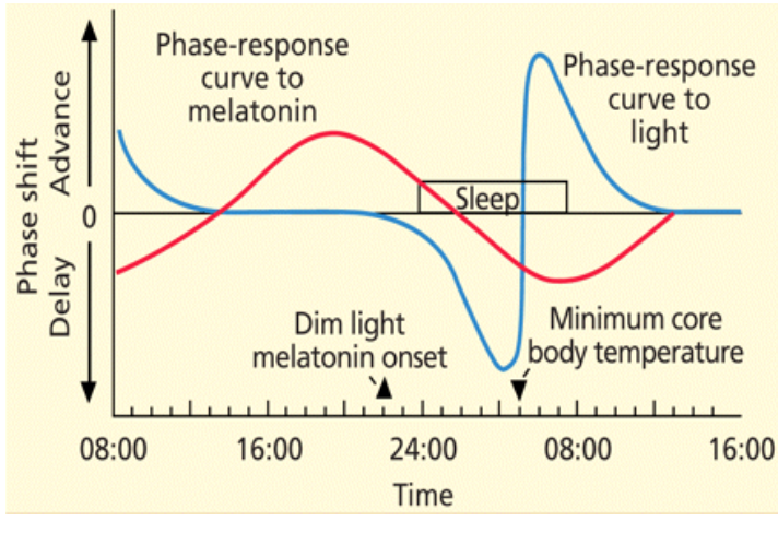
Pictured is what is known as the human phase response curve to light and melatonin. Meaning that it maps our level of responsiveness to one or the other while showing just how exactly it will affect our circadian rhythm: delay means that you will sleep and wake up later, while an advance means that you will sleep and wake up earlier.
By using this curve (while also accounting for individual differences, such as chronotype) we can manipulate our circadian rhythm to be better for us. See the next section for a breakdown on changing your rhythm.
How can I curb the effects of jet lag when travelling?
According to the phase response curve to light, in order to delay our clock we need to get light late in the day (i.e. evening), whereas in order to advance our clock we need to get light early in the day (i.e. morning). The crossover point between delays and advances is around our body temperature minimum.
Step 1: Estimate your body temperature minimum by finding the mid-point of your normal sleep cycle (i.e. if you sleep from 12AM to 8AM, your midsleep will be 4AM, which can be used as an approximation of your body temp minimum).
Step 2: Decide if you want to delay or advance your clock. General rule: if you are traveling east, it will be easier to advance your rhythms since you want to get up earlier than usual. If you are traveling west, it will be easier to delay your rhythms since you want to get up later than usual.
Step 3: Convert your body time to the new time zone. If your body temp min is 4AM in Vancouver, that means it is occurring at 6AM Toronto time (4AM PST = 6AM ET), and since T.O. is east of here, we want to advance our rhythms. Therefore we want to avoid light prior to 6AM (since this would cause delays) and seek light after 6AM (which will lead to advances).
The circadian clock can shift ~1 hour per day, so to move two time zones, it should take 2 days to adjust in this example. The benefit of developing a schedule like this is to minimize the amount of jetlag.
If you need to advance your rhythms and you get light during the phase delay portion of your day, then you are actually pushing yourself the wrong direction! This will lead to increased duration of jetlag. Maybe not such a big deal traveling to Toronto, but for overseas trips this could be no fun.
Below are some of the factors that contribute to how you experience jetlag and what you can do to mitigate its effects on your circadian rhythm.
- Shift magnitude: the larger the time difference, the longer it will take for re-entrainment to occur (for you to adjust to the new time zone). Humans and hamsters travelling eastwards will shift about ~1 hour per day.
- Direction of travel: re-entrainment occurs more quickly when travelling westward, with respect to the shape of your phase response curve and chronotype. The human circadian cycle tends to exceed 24h just a little, thus the natural delay allows us to entrain more easily to a western time zone.
- Timing of light exposure: in a new time zone, light exposure at certain times will either accelerate or impede re-entrainment. The right times for acceleration and the wrong times leading to impediment depend on the direction of travel and can be induced with the use of the phase response curve.
- Parameters of the light stimulus: light parameters include amplitude (intensity) and length (colour) of waves. The brighter the light, the bigger the shift, with blue light (480nm) being the most effective.
What is a “body clock”?
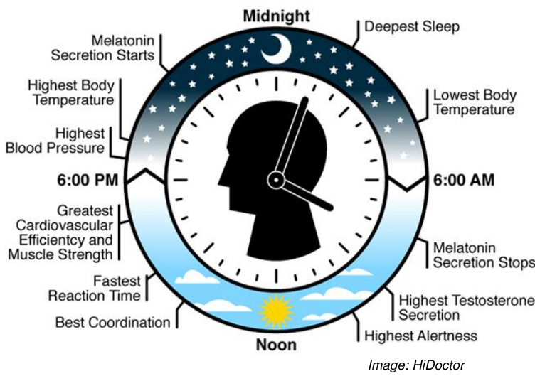
A “body clock” is a simplified pictorial representation of some of the major bodily functions that change depending on the time of day, and is mapped out in relation to what is considered to be a normal chronotype. As you can see, our endocrine and digestive systems are closely tied with our aforementioned ‘master clock’. This is because many of our other biological clocks (called peripheral clocks), such as the ones that dictate our gut and hormonal systems, must stay in sync with our master clock in order for things to run smoothly.
In the absence of light, many of our digestive hormones are not released properly and can result in gastrointestinal disorders, while constant disruption of our natural sleep cycle can interfere with stomach acid secretion, which may lead to Irritable Bowel Syndrome, gastrointestinal reflux, or ulcers. Many hormones also secrete on a 24hr cycle: cortisol (associated with stress), for example, shows a clear rise in the morning and fall in the evening.
This is why shiftwork is very dangerous, as it induces internal desynchrony between your master clock and major organs, resulting in many health problems. Cognitive performance also varies throughout the day depending on how long you’ve been awake and what time of day it is, but typically most tasks are best performed during the second half of the day, as it peaks with body temperature.
References
- Allada, R. (2003). Circadian clocks: A tale of two feedback loops. Cell, 112(3), 284-286. https://doi.org/10.1016/S0092-8674(03)00076-X
- Brown, G. M. (1994). Light, melatonin and the sleep-wake cycle, 345–353.
- Czeisler, C. A., & Gooley, J. (2007). Sleep and circadian rhythms in humans. Cold Spring Harbor Symposia on Quantitative Biology, 72, 579–597. https://doi.org/10.1101/sqb.2007.72.064
- Dunican, I. C., & Caldwell, J. (2015). Managing sleep and jet lag for optimal performance (November).
- Emens, J. S. (2017). Circadian rhythms: The price of electric light. Current Biology. https://doi.org/10.1016/j.cub.2017.01.014
- Ferrante, A., Gellerman, D., Ay, A., Woods, K. P., Filipowicz, A. M., Jain, K., … Ingram, K. K. (2015). Diurnal preference predicts phase differences in expression of human peripheral circadian clock genes. Journal of Circadian Rhythms, 13, 1–7. https://doi.org/10.5334/jcr.ae
- Fonken, L. K., & Nelson, R. J. (2014). The effects of light at night on circadian clocks and metabolism. Endocrine Reviews, 35(4), 648–670. https://doi.org/10.1210/er.2013-1051
- Gamble, K. L., & Young, M. E. (2015). Circadian biology: The early bird catches the morning shift. Current Biology, 25(7), R269–R271. https://doi.org/10.1016/j.cub.2015.02.004
- Glickman, G., Byrne, B., Pineda, C., Hauck, W. W., & Brainard, G. C. (2006). Light therapy for seasonal affective disorder with blue narrow-band light-emitting diodes (LEDs). Biological Psychiatry, 59(6), 502–507. https://doi.org/10.1016/j.biopsych.2005.07.006
- Graham, D., & Wong, K. Y. (2016). Photosensitive retinal ganglion cells (ipRGCs). WebVision, 1–47.
- Harmer, S. L., Panda, S., & Kay, S. A. (2001). Molecular bases of circadian rhythms.
- Kelleher, F. C., Rao, A., & Maguire, A. (2014). Circadian molecular clocks and cancer. Cancer Letters, 342(1), 9–18. https://doi.org/10.1016/j.canlet.2013.09.040
- Lockley, S. W., Arendt, J., & Skene, D. J. (2007). Visual impairment and circadian rhythm disorders. Dialogues in Clinical Neuroscience, 9(3), 301–314. https://doi.org/10.31887/DCNS.2007.9.3/slockley
- Morf, J., & Schibler, U. (2013). Gatekeepers of circadian clocks. Cell Cycle, 12(4), 539–40. https://doi.org/10.4161/cc.23670
- Roenneberg, T., & Merrow, M. (2007). Entrainment of the human circadian clock. Cold Spring Harbor Symposia on Quantitative Biology, 72, 293–299. https://doi.org/10.1101/sqb.2007.72.043
- Simons, S. S. H., Beijers, R., Cillessen, A. H. N., & Weerth, C. de. (2015). Development of the cortisol circadian rhythm in the light of stress early in life. Psychoneuroendocrinology, 62, 292–300. https://doi.org/10.1016/j.psyneuen.2015.08.024
- Toh, K. L. (2008). Basic science review on circadian rhythm biology and circadian sleep disorders. Annals of the Academy of Medicine Singapore, 37(8), 662–668.
- Vetter, C., Fischer, D., Matera, J. L., & Roenneberg, T. (2015). Aligning work and circadian time in shift workers improves sleep and reduces circadian disruption. Current Biology (Vol. 25). https://doi.org/10.1016/j.cub.2015.01.064
- West, A. C., & Bechtold, D. A. (2015). The cost of circadian desynchrony: Evidence, insights and open questions. BioEssays, 37(7), 777–788. https://doi.org/10.1002/bies.201400173
- Wright, K. P., McHill, A. W., Birks, B. R., Griffin, B. R., Rusterholz, T., & Chinoy, E. D. (2013). Entrainment of the human circadian clock to the natural light-dark cycle. Current Biology (Vol. 23). https://doi.org/10.1016/j.cub.2013.06.039